Intraoperative Imaging
Intraoperative imaging and monitoring provides surgeons with real-time physiologocal data that can help guide surgery, and can improve post-operative outcomes and long-term quality of life. In this context, optical imaging offers very viable, portable, inexpensive and real-time solutions that are sensitive to several physiological parameters of interest. Research in this area involves the development of new methods that can address fairly unique needs of intraoperative imaging, for applications that include quantitative imaging of blood flow during neurosurgeries, aneurysm clippings & carotid endarterectomies, and intraoperative detection of tumor margins with fluorescence.
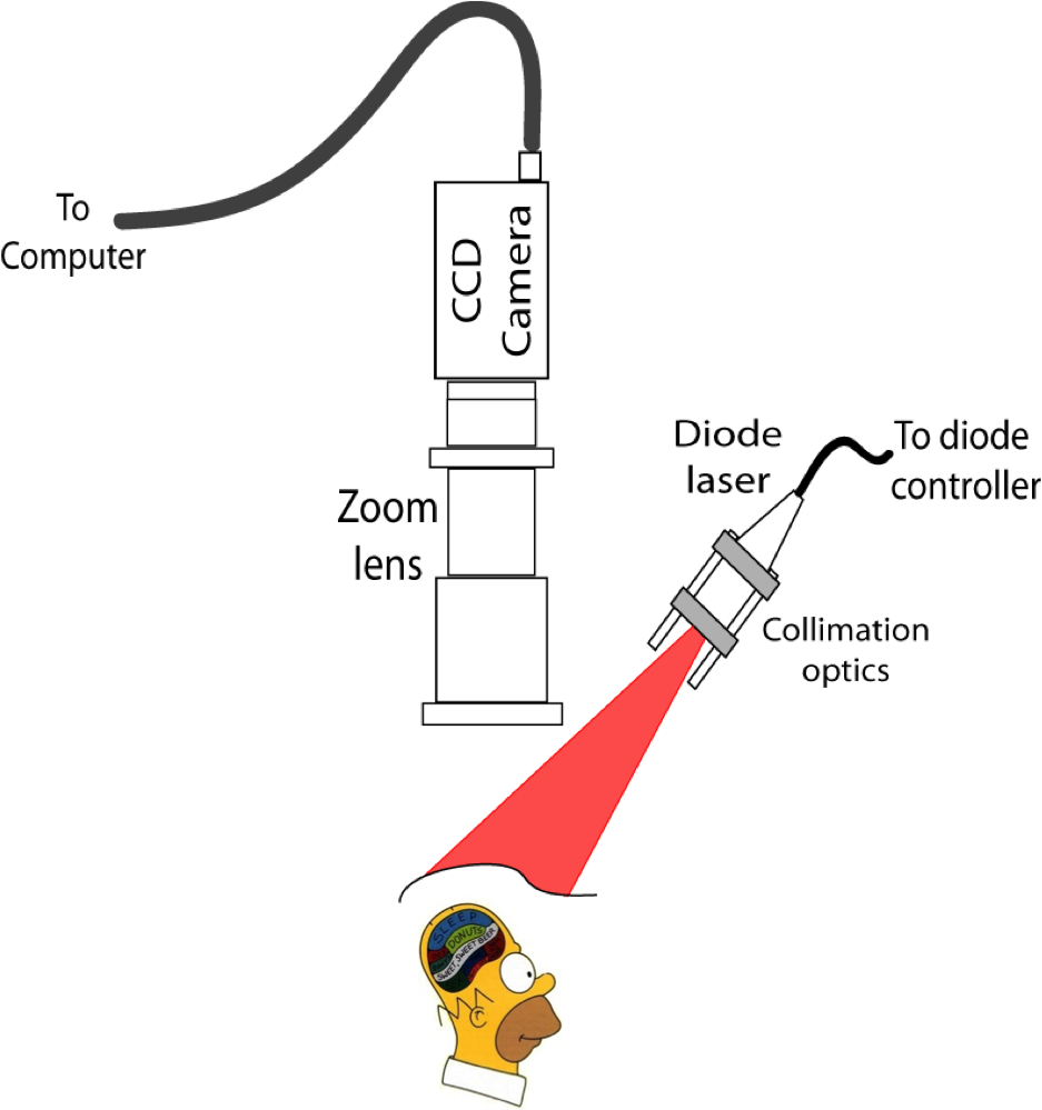
See also Laser Speckle Contrast Imaging for more information.
Back to Research Topics
Relevant Publications
Holt D, Parthasarathy AB, Okusanya O, Keating J, Venegas O, Yodh AG, Deshpande C, Karakousis G, Madajewski B, Durham A, Nie S, and Singhal S (2015). “Intraoperative near-infrared fluorescence imaging and spectroscopy identifies residual tumor cells in wounds”, Journal of Biomedical Optics, 20 (7), 076002, PMID: 26160347 PMCID: PMC4497968.(PDF)
Parthasarathy AB, Weber EL, Richards LM, Fox DJ, and Dunn AK, (2010) “Laser Speckle Contrast Imaging of Cerebral Blood Flow in humans during neurosurgery: A pilot clinical study”, Journal of Biomedical Optics, 15 (6), 066030, Pubmed PMID: 21198204.(PDF)