Laser Speckle Contrast Imaging
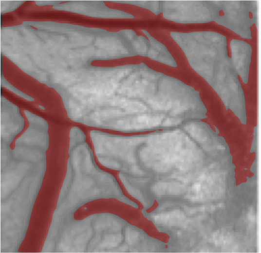
Laser Speckle Contrast Imaging (LSCI), is a camera-based optical technology, that is particularly useful for imaging cerebral blood flow using intrinsic contrast. LSCI’s relatively simple instrumentation offers superior spatial and temporal resolution; the technique has been widely used to image CBF dynamics during ischemia and functional activation in rat and mouse brain. A typical LSCI instrument illuminates the sample using slightly divergent light from a single mode diode laser (operating in the visible or near infrared (NIR)). The backscattered light that has interacted with the tissue is collected through an imaging system and recorded on the camera sensor (typically 8 bit). The data is stored in a computer, wherein raw intensity (speckle) images are converted to images of cerebral blood flow (speckle contrast); (See Laser speckle contrast imaging in biomedical optics, by Boas & Dunn, Journal of Biomedical Optics (2010) for a comprehensive review of LSCI)
Briefly, laser speckle is the random interference pattern produced by coherent addition of laser light beams that has backscattered from a sample, along trajectories with slightly different path lengths. Similar to Diffuse Correlation Spectroscopy, movements of particles in the sample (i.e., red blood cells), impress spatial and temporal fluctuations onto the speckle pattern. In the spatial domain, these fluctuations are manifest as local blurring (or decorrelation) in the image. LSCI quantifies blood flow by calculating the local speckle contrast - the square root of normalized variance of local intensity (K= σ/I), over a moving window (usually 7×7 pixels); here, σ is the standard deviation of the intensities in the 7x7 pixel window, and I is the mean of intensity in the 7x7 pixel window. In the resulting speckle contrast image, low contrast values (e.g., in a major vessel) is a reflection of increased blurring of speckles due to large RBC speeds, and hence reduced variance in intensities.
Current research in LSCI is focused on improving the quantitative accuracy of blood flow measurements, e.g., using Multi Exposure Speckle Imaging, (see Publications below), adapting LSCI for intraoperative imaging (see Intraoperative Imaging), miniaturization of instrumentation, real-time flow computation, and 3-D imaging.
Back to Research Topics
Relevant Publications
S. M. Shams Kazmi, Parthasarthy AB, Song N. E, Jones T.A, and Dunn AK (2013), “Chronic Imaging of Cortical Blood Flow using Multi-Exposure Speckle Imaging”, Journal of Cerebral Blood Flow & Metabolism, 33 (6), 798-808, PMID: 23571277 PMCID: PMC3677120.(PDF)
Parthasarathy AB, Weber EL, Richards LM, Fox DJ, and Dunn AK, (2010) “Laser Speckle Contrast Imaging of Cerebral Blood Flow in humans during neurosurgery: A pilot clinical study”, Journal of Biomedical Optics, 15 (6), 066030, Pubmed PMID: 21198204.(PDF)
Parthasarathy AB, S.M. Shams Kazmi, and Dunn AK (2010). “Quantitative imaging of ischemic stroke through thinned skull in mice with Multi Exposure Speckle Imaging”, Biomedical Optics Express, 1 (1), 246-259, PMID: 21258462 PMCID: PMC3005179.(PDF)
Zaman RT, Parthasarathy AB, Vargas G, Chen B, Dunn AK, Rylander III HG, and Welch AJ (2009). “Perfusion in hamster skin treated with glycerol”, Lasers in Surgery and Medicine, 41 (7), 492-503, PMID: 19670326.(PDF)
Parthasarathy AB, Tom WJ, Gopal A, Zhang XJ, and Dunn AK (2008). “Robust flow measurement with multi-exposure speckle imaging”, Optics Express, 16 (3), 1975-1989, PMID: 18542277.(PDF)